


If you have been told you need a procedure to treat your kidney stone and the word “ureteroscopy” or “fURS” has come up, this article is for you. We want to give you a clear, honest picture of what that procedure actually involves — including risks that are rarely discussed in a rushed clinic appointment — so you can have an informed conversation with your doctor about whether a simpler, non-invasive option might suit you better.
What Is Flexible Ureteroscopy (fURS)?
Flexible ureteroscopy — often called fURS or RIRS (retrograde intrarenal surgery) — is a procedure in which a thin, flexible tube with a camera at its tip is passed through your urethra, up through your bladder, and into the ureter (the tube connecting your kidney to your bladder), all the way into the kidney itself. A laser fibre is then threaded down the same tube to break the stone into smaller pieces or dust.
The procedure requires general or spinal anaesthesia. It is increasingly popular among urologists because it can achieve high stone-clearance rates. But “popular” and “risk-free” are not the same thing, and what happens to your urinary tract in the process deserves careful attention.
The Two Types of Damage fURS Can Cause
- Mechanical / Instrumentation DamageThe flexible ureteroscope is a remarkably delicate instrument — and so is your ureter. Getting the scope safely into the kidney requires passing it through a narrow tube that was never designed to accommodate surgical instruments. This is why mechanical injuries to the ureter are among the most commonly reported complications of fURS.Among 1,571 procedures reviewed in one large study, the primary intraoperative complications were bleeding (2.5%) and mucosal injury (2.3%), while ureteral perforation — requiring immediate stent insertion — was also observed. These figures represent the routine surgical experience, not exceptional cases.The access sheath — a plastic sleeve inserted into the ureter to allow the scope to pass in and out — is itself a source of harm. The ureteral access sheath is extensively used in fURS due to its benefits for stone clearance, but it may cause damage to the ureteral wall, and its insertion is recognised as a leading cause of ureteral injury.In cases of impacted stones — stones that have been stuck in the ureter for some time — the risks are substantially higher. Patients with impacted stones face increased risks of surgical failure, ureteral rupture, and urosepsis, and ureteral stricture is a common complication after fURS in these cases, with studies reporting up to a 24% chance of postoperative ureteral stricture. A ureteral stricture means scarring that narrows the ureter, which can obstruct urine flow from the kidney and lead to long-term kidney damage if untreated.
- 13.3%Overall complication rate in one large fURS study (1,571 cases)
- Up to 24% Risk of ureteral stricture with impacted stones
- 1–4% Rate of post-ureteroscopy ureteral stricture overall
- Thermal (Heat) Damage from the Laser
The laser used to break stones during fURS generates significant heat — and the delicate lining of your ureter is in the firing line. This is one of the most underappreciated risks of the procedure.
Once the temperature exceeds the threshold of 43 °C, it can lead to cell damage, protein coagulation, and tissue injury, which subsequently progresses to scar formation and ureteral stricture. When the temperature reaches 56 °C, it takes only one second to cause thermal damage to the tissue — so thermal injury to the ureter can easily go unnoticed during lithotripsy.
In plain terms: the laser heats the fluid inside your ureter. If the heat builds up even briefly, the delicate lining of the ureter can be permanently scarred. The surgeon may not know this is happening — the damage only becomes visible weeks or months later when the ureter starts to narrow.
Research confirms this is a real clinical problem. It has been reported that post-ureteroscopy ureteral stricture can be as high as 1–4%, and the potential cause of stricture is more likely to be the direct thermal injury towards the ureteral wall rather than acoustic or photonic energy.
The choice of laser also matters. A large retrospective database analysis of 6,726 patients who underwent ureteroscopic laser lithotripsy found that the risk of stricture formation was 4.6% in the thulium fiber laser group versus 2.4% in the holmium laser group — nearly double the risk with the newer laser technology. This is particularly relevant because the newer thulium fiber laser is being rapidly adopted in many centres precisely because it fragments stones faster — but at the cost of greater thermal output.
“When the temperature reaches 56 °C, it takes only one second to cause thermal damage to ureteral tissue — damage the surgeon cannot see and the patient will not feel until weeks later.”
Why You Need a Stent Before the Procedure Even Begins
Here is something many patients are surprised to discover: in many cases, fURS cannot simply be performed on the day you arrive for surgery. Your ureter may be too narrow for the scope to pass safely, and the surgeon needs to stretch — or “dilate” — it first.
The standard way to do this is to insert a JJ stent (also called a double-J stent) in a separate procedure, wait 1–4 weeks for the ureter to passively stretch around it, and only then perform the fURS. Preoperative stenting is frequently used to allow passive ureteral dilatation, which is supposed to facilitate the passage of a flexible ureteroscope or ureteral access sheath.
Patients who were pre-stented for any reason demonstrated significantly improved ureteroscopic access and stone-free rates, with pre-stenting associated with lower need for postoperative stenting and fewer complications.
What this means in practice: before your kidney stone is even touched, you may need to spend two to four weeks with a stent inside you — enduring all the discomfort that comes with it (described in the next section) — simply to make the main procedure possible. For many patients, this is an unwelcome surprise that was not clearly explained before they agreed to the surgery.
Living with a JJ Stent: The Reality
A JJ stent is a soft plastic tube curled at both ends — one curl sits in your kidney, the other in your bladder — to keep the ureter open and allow urine to drain. It sounds simple. The reality of living with one is considerably less so.
More than 80% of patients with a ureteral stent present with associated symptoms, including pain, lower urinary tract symptoms, and sexual dysfunction, leading to an important drop in quality of life.
Among the most common symptoms associated with ureteral catheters are those associated with lower urinary tract symptoms (LUTS), such as dysuria, urinary urgency, increased frequency, the feeling of incomplete emptying of the bladder, urinary incontinence, or even urinary tract infections. These are not rare side effects — they are the expected experience for the majority of patients.
- Flank and Back Pain
Over 80% of stented patients experience pain, often in the kidney area, that disturbs sleep and daily activity. - Urinary Urgency & Frequency
The bladder end of the stent constantly irritates the bladder wall, causing overactive bladder-like symptoms. - Blood in Urine
Haematuria is a common occurrence as the stent rubs against the internal lining of the urinary tract. - Infection Risk
The stent is a foreign body and a magnet for bacteria. Repeated infections, and in rare cases sepsis, can occur. - Sleep Disruption
Studies report that up to 77% of patients have their sleep interrupted due to stent-related pain and urinary symptoms. - Sexual Dysfunction
Stent-related discomfort significantly impacts sexual life in both men and women, including dyspareunia and reduced libido.
A 2023 study found that among 200 patients with ureteral stents, urgency was reported in 90.5% of cases, dysuria in 50.5%, and haematuria in 40%, with symptoms negatively impacting both work performance and sexual health. Significant improvements were only observed after stent removal.
Now consider the timeline for a patient undergoing fURS: a stent is placed to pre-dilate the ureter (weeks 1–4), then the fURS procedure is performed under general anaesthesia, and then a further stent is placed post-operatively to let the ureter heal (weeks 4–8). That can mean two to three months of stent-related symptoms — all for a procedure to treat a stone that was causing you pain in the first place.
The Stent Timeline Many Patients Don’t Expect
- Week 1–4: Pre-operative JJ stent placed to dilate the ureter. Patient experiences urinary symptoms, pain, and possible infections.
- Week 4–5: fURS procedure under general anaesthesia. Laser fired inside the ureter. Risk of mechanical and thermal injury.
- Week 5–8+: Post-operative stent placed to protect the ureter as it heals. Patient experiences the same cycle of symptoms again.
- Then: Stent removed in a further cystoscopic procedure — itself requiring local or general anaesthesia.
There Is an Alternative: ESWL
Extracorporeal shock wave lithotripsy (ESWL) is a fundamentally different approach to the same problem. Instead of sending instruments inside your body, ESWL uses focused shock waves from a machine outside your body to break the stone — with no incisions, no scope, and typically no stent.
ESWL is the only truly non-invasive outpatient surgical procedure for renal and ureteral calculi. Its advantages compared to ureteroscopy include: it is less invasive and very well tolerated; it carries a much lower complication rate; it is an inherently safer procedure; it requires no double-J stents in the majority of cases; no pre-stenting is needed; and there is no need for ureteral dilation or ureteral access sheaths.
This means that for suitable patients, ESWL can offer stone treatment without the weeks of pre-operative stenting, without general anaesthesia in most cases, without a laser being fired inside the ureter, and without the risk of ureteral scarring and stricture that accompanies scope surgery.
ESWL can be performed with intravenous sedation or even local anaesthesia, unlike ureteroscopy which involves general or spinal anaesthesia. Studies have shown that stents have no benefit and are therefore not needed for patients undergoing ESWL, especially if the stone is small.
ESWL is most effective for stones up to about 2 cm in size, located in the kidney or upper ureter. Stone composition matters too — brittle, less dense stones respond best. Recent data shows initial success rates of around 69–93% depending on stone characteristics and the technology used, with repeat sessions available if needed.
A Simpler Comparison
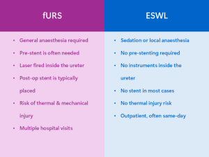
What to Ask Your Doctor
If your surgeon has recommended fURS, it does not mean it is your only option or the wrong option for you. Every case is different. But you deserve a full picture. Here are some questions worth raising at your next appointment:
Questions Worth Asking
- Am I a candidate for ESWL? Why or why not?
- Will I need a pre-operative stent before my ureteroscopy? For how long?
- What is the risk of ureteral injury or stricture with this procedure?
- What type of laser will be used, and what are the associated thermal risks?
- Will I need a post-operative stent, and how long will it stay in?
- What is the total number of procedures and anaesthetics likely to be involved?